Scanning X-ray Microscopy with scattering (SAXS/WAXS) and absorption contrast as a diagnostic tool for biotissues and pathologies
Quantitative and multi-length scale structural imaging of biotissues is performed through the SAXS/WAXS/absorption contrast detection. Crystalline order modifications at the atomic and nano scale are related to physio-pathologic conditions of the collagenic and mineral components, as to fundamental biomineralization processes.
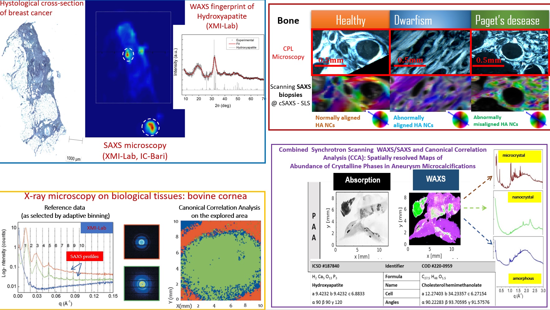
Scanning SAXS/WAXS experiments were performed by using synchrotron radiation and/or laboratory sources on: pathological corneal tissues affected by keratoconus; bone tissues affected by dwarfism and Paget’s disease; biopsies of aneurysms; tumor tissues with microcalcifications. Many types of cancer (prostate; breast; thyroid; glioblastoma) involve (micro)calcifications, which therefore represent the marker of a benign or malignant neoplasm. Several studies have brought to attention the significance of crystallographic data with respect to diagnosis of malignancy. Hence it is crucial to assess the origin, composition and crystalline structure of calcifications, and if these change from benign to malignant lesions, opening new important diagnostic and therapeutic perspectives in tumors with or without calcifications. SAXS/WAXS microscopy allows the localization and the identification of the different crystalline phases, as well as other components within the investigated tissue such as collagen and myofilament, useful for an overall assessment of the structural modifications related either to pathologies like tumors featuring evident microcalcifications, or to pathologies mainly involving alterations of the nanoscale collagen structure, such as keratoconus in the cornea.
– T Sibillano, L De Caro, F Scattarella, G Scarcelli, D Siliqi, D Altamura, M Liebi, M Ladisa, O Bunk, C Giannini, Interfibrillar packing of bovine cornea by table-top and synchrotron scanning SAXS microscopy, J Appl Crystallogr . 2016 Jul 14;49(Pt 4):1231-1239.
– R. Vanna, C. Morasso, B. Marcinn, F. Piccotti, E. Torti, D. Altamura, S. Albasini, M. Agozzino, L. Villani, L. Sorrentino, O. Bunk, F. Leporati, C. Giannini, and F. Corsi. Raman Spectroscopy Reveals That Biochemical Composition of Breast Microcalcifications Correlates with Histopathologic Features. Cancer Res; 80(8) April 15, 2020, 1762-1772.
– Giannini, M. Ladisa, V. Lutz-Bueno, A. Terzi, M. Ramella, L. Fusaro, D. Altamura, D. Siliqi, T. Sibillano, A. Diaz, F. Boccafoschi and O. Bunk. X-ray scanning microscopies of microcalcifications in abdominal aortic and popliteal artery aneurysms. IUCrJ (2019). 6, 267–276.
– C. Giannini, D. Siliqi, O. Bunk, A. Beraudi, M. Ladisa, D. Altamura, S. Stea & F. Baruffaldi. Correlative Light and Scanning X-Ray Scattering Microscopy of Healthy and Pathologic Human Bone Sections. SCIENTIFIC REPORTS, 2012, 2 : 435, DOI: 10.1038/srep00435.